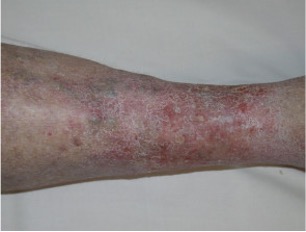
Vein disease can affect the skin as well as the veins. Longstanding venous pressure may contribute to inflammation, staining and tissue changes. These findings deserve examination because infection, arterial disease and other skin conditions may look similar or coexist. A photograph can help document a change, but cannot confirm the diagnosis.
Why is the skin around my ankles turning brown?
Brown staining around the lower leg or ankle can occur when venous pressure allows blood components to move into surrounding tissues, leaving pigment. It is one possible explanation, not the only cause of discolouration. New or progressive staining, particularly with swelling or itching, is worth assessing.
Can varicose veins cause eczema?
Venous disease can contribute to venous eczema: itchy, inflamed skin, often on the lower legs. The skin may be dry, scaly, cracked or weeping. Managing the skin alone may not address an underlying circulation problem, so the assessment should consider both.
How is venous eczema different from other types of eczema?
Venous eczema is associated with impaired venous return and may occur alongside swelling or brown staining. Other eczema patterns and contact allergy can overlap with it. A clinician considers the distribution, history and other leg findings; a red or itchy patch should not automatically be labelled venous eczema.
What are the small fan-shaped veins around my ankle?
A fan-like cluster of small veins around the ankle may be called corona phlebectatica. Certain patterns are associated with more advanced venous disease and merit assessment. Not every visible ankle vein has the same significance, and the finding does not mean an ulcer is inevitable.
What does lipodermatosclerosis mean?
Lipodermatosclerosis is inflammation and hardening of the skin and the fatty tissue beneath it, associated with chronic venous disease. The lower leg may feel tender, firm or tight. Examination helps distinguish it from other causes of an inflamed leg.
What is the difference between venous eczema and lipodermatosclerosis?
Venous eczema describes an inflammatory skin rash. Lipodermatosclerosis involves hardening of deeper tissues as well. They can occur together, and neither should be diagnosed from a photograph alone.
Could a sore near my ankle be a venous leg ulcer?
A wound near the ankle may be a venous ulcer, but location alone is not enough. Assessment considers the wound, venous circulation and arterial blood supply. Care may combine wound treatment, appropriately selected compression and treatment of relevant venous reflux. A persistent wound should not simply be covered and forgotten.
When should a slow-healing leg wound be assessed?
Arrange prompt assessment of a wound that is not healing or repeatedly breaks down; do not wait for it to become large. Increasing pain, spreading redness, discharge or fever require more urgent advice. A wound that does not respond as expected needs its diagnosis and treatment reviewed, including possible non-venous causes.
For your next conversation
Help the clinician see what has changed
- Bring a dated record of when the skin change or wound appeared and any previous episodes.
- List creams, dressings, adhesives and stockings used, including any irritation they caused.
- Ask whether both arterial and venous circulation need assessment before compression is started or increased.
General information to support a discussion with your treating clinician. It does not replace individual assessment or discharge instructions. In an emergency in Australia, call 000.