Conditions
What is chronic venous disease?
Chronic venous disease is the umbrella term for leg veins that no longer return blood to the heart efficiently, usually because the one-way valves inside them have weakened. It covers both what you can see, such as varicose veins or eczema-like skin changes, and what you can feel, such as aching, heaviness, throbbing or restless legs that build towards the end of the day. It is common, it develops gradually, and it sits on a spectrum from minor to serious.
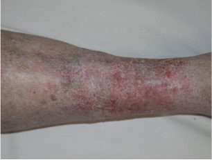
What it is
Veins in the legs have a hard job: they carry blood upward, against gravity, back to the heart. Two things make that possible. The calf muscle acts as a pump, squeezing blood up the leg with every step, and a series of one-way valves inside the veins close between squeezes so the blood cannot fall back down.
Chronic venous disease is what develops when those valves weaken and start to leak. Each time the calf relaxes, some blood slips back down the vein. Over months and years the pressure in the leg veins rises. Veins stretch and become tortuous, fluid leaks into the surrounding tissue, and the skin of the lower leg is slowly affected by the sustained pressure.
The term covers both halves of the picture. The visible part is what people describe as varicose veins, spider veins or eczema on the legs. The functional part is what people feel: aching, heaviness, throbbing and restlessness that build through the day and ease with the legs up.
It is common. Estimates in the original CVLM article suggested that up to 60 per cent of adults may have some form of venous disease, with roughly one in three women and one in five men affected. Women are affected more often, and the likelihood rises with age, pregnancy, family history, and years spent standing.
It also sits on a spectrum. At one end are small surface veins and mild end-of-day symptoms. At the other are large, ropey varicose veins, lasting skin changes and venous ulcers. Where you sit on that spectrum cannot be judged from the surface.
Signs to look for
Symptoms are often gradual enough that people adapt around them without noticing. Common ones include:
- Aching, dragging or heavy legs, worse after standing and later in the day
- Throbbing, burning or cramping in the calves, particularly at night
- Ankle or lower-leg swelling that varies through the day
- An urge to move the legs at rest, or difficulty settling at night
- Visible veins: fine surface veins, or raised, twisting veins beneath the skin
- Itching or a rash on the lower leg
Skin changes suggest the pressure has been present for some time. The medical terms you may see in a report include:
- Venous eczema: dry, itchy, red or scaly skin, usually around the ankle and shin
- Corona phlebectatica: a fan of fine purple veins around the ankle and the side of the foot
- Lipodermatosclerosis: hardening and tightening of the skin and tissue of the lower leg, often with brown discolouration, sometimes giving the leg an inverted-bottle shape
- Atrophie blanche: small, smooth, white patches of scar-like skin, usually near the ankle, which can be fragile

A sore near the ankle that is slow to heal, or that keeps reopening, is the most advanced sign and is covered on the venous leg ulcers page.
It is not unusual for people to play down their symptoms because they assume the only fix is surgery. That is rarely the case now; most vein treatment is performed in the clinic under local anaesthetic, without a hospital stay. If any of the signs above are familiar, arrange an assessment.
What we check
Your first appointment has three parts and they happen on the same day.
First, a conversation. What you have noticed, how long it has been present, what makes it better or worse, your medical history, medications, previous vein treatment, and any family history of vein disease or clots.
Second, an examination of both legs: the veins, the skin, and any swelling.
Third, a duplex ultrasound of the leg veins, performed on site. This is the test that actually diagnoses chronic venous disease. It shows the direction of blood flow in each vein and identifies which valves are leaking and where. Two people with identical-looking legs can have completely different findings underneath, which is why the scan comes before any discussion of treatment.
You are then shown what was found, how it relates to your symptoms, and what the options are. Everything is supplied in writing so you can consider it at home.
Treatment options
Where treatment is clinically indicated, the options depend on which veins are involved. In broad terms:
- Compression and movement manage symptoms and are part of every plan.
- Endovenous laser ablation (EVLA) closes a leaking main vein from the inside, under local anaesthetic.
- Ultrasound-guided sclerotherapy treats veins that are not visible on the surface, using ultrasound to guide the injection.
- Microsclerotherapy treats fine surface veins.
The treatment pages explain each in more detail, including what recovery involves. Some presentations are better managed conservatively or referred on, and that is discussed honestly.
A Medicare rebate applies where treatment is clinically indicated and eligibility criteria are met.
When to be assessed
Chronic venous disease is progressive, and the skin changes it causes become harder to reverse the longer the pressure persists. You do not need a threshold of discomfort before asking. Persistent or changing leg symptoms, swelling on most days, itching or discolouration on the lower leg, or a wound near the ankle that is slow to heal are each a reason to arrange an assessment.
Seek urgent care rather than a routine appointment for sudden swelling or pain in one leg, chest pain or breathlessness, or bleeding from a vein that does not stop with firm pressure. Call 000 in an emergency.
Canberra Vein & Laser Medicine is at The Woden Suites, Level 3, Francis Chambers, 40 Corinna Street, Phillip ACT 2606. Book online or call (02) 5119 3136.
FAQ
Questions people ask
- What is chronic venous disease?
- Chronic venous disease is a long-term condition in which the leg veins struggle to return blood to the heart, most often because their one-way valves leak. Blood pools, pressure rises, and that shows up as varicose veins, aching, heaviness, swelling, skin changes or, in advanced cases, ulcers. It is diagnosed with a history, examination and duplex ultrasound.
- Is chronic venous disease serious?
- It ranges widely. Many people have mild symptoms and small visible veins for years. At the other end, untreated venous pressure can lead to skin hardening, discolouration, eczema and leg ulcers that are slow to heal. The condition is progressive, so persistent or changing symptoms are worth assessing rather than waiting for them to worsen.
- What is the difference between chronic venous disease and varicose veins?
- Varicose veins are one visible sign of chronic venous disease, not the whole condition. Chronic venous disease describes the underlying valve failure and raised pressure; varicose veins, spider veins, swelling, skin changes and ulcers are the ways it can present. You can have significant venous disease with few visible veins, and visible veins with little underlying disease.
- How is chronic venous disease diagnosed?
- With a duplex ultrasound of the leg veins, alongside a history and examination. The scan shows the direction of blood flow and identifies which valves are leaking and which veins are affected. At CVLM the ultrasound is performed on site during your first appointment, so findings are explained the same day.
- Can chronic venous disease be reversed?
- Damaged valves do not repair themselves, but the veins responsible can often be treated so that blood is redirected through healthy veins, and symptoms and skin changes frequently improve as a result. Compression and movement help manage symptoms. What is appropriate depends on your ultrasound findings, so arrange an assessment before assuming either way.